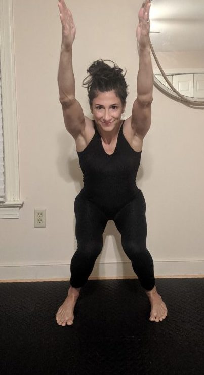
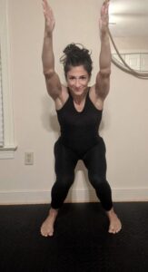
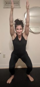
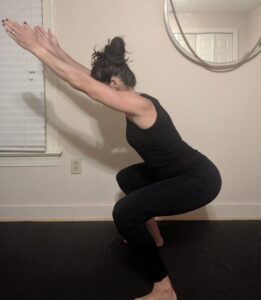
After getting familiar with performing an overhead squat assessment (OHSA) and noting any signs of imbalance or dysfunction, you’ll have to know what to do with that information to be of any help to your fitness clients. Start from the ground up, since so much dysfunction is rooted in the lower extremities, makes good, logical sense.
Specific problems with the feet are left out of this assessment because they warrant their own category of dysfunction.
(Bear in mind, when programming corrective exercise, you will want to start with any area that is causing a problem or pain first. In the absence of those conditions, start with correcting LED)
Signs of LED and What They Mean
- Knees bow in—Functional knee valgus (knees are closer than hips and feet) or even a knee-wobble on the descent or turn-around can be a sign of LED and/or Lumbo Pelvic Hip Complex Dysfunction (LPHCD). Look for this sign on the transition of eccentric to concentric motion. This pattern is an indication of excessive femoral internal rotation and excessive tibial external rotation. Femoral internal rotation points to tight tensor fascia latae (TFL), vastus lateralis and gluteus minimus. The anterior adductors are also short. Expect the opposing femora external rotators (piriformis, gluteus maximus and glute medius, etc) to be weak.
Furthermore, tibial external rotators will be overactive with this pattern (as knees bow in, the tibia is turning out). This includes biceps femoris short head, the lateral head of gastrocnemius in addition to the same femoral internal rotators mentioned above. The lesser-known tibial internal rotators, gracilis, popliteus, semi-tendinosus and -membranosus and the Vastus Medialus Obliquus (VMO), are all underperforming.
- Feet flatten—Excessive eversion signals short fibularis and lateral gastroc, in addition to short plantarflexors soleus, gastroc, and digital flexors. Expect the opposing muscle groups that perform inversion and dorsiflexion, anterior and posterior tibialis to be weak. (Even though post tib plantarflexes, it also inverts the foot, so it’s weakness contributes to excessive eversion)
- Feet turn out—When the knees are directed straight ahead but the feet are externally rotated (tibial external rotation), both LED and LPHCD are present. This dysfunction is a result of lack of dorsiflexion (plantarflexors are tight) in addition to compensatory tibial external rotation. The tibial external rotators are short and tibial internal rotators are weak.
- Excessive forward lean—This compensatory pattern is a sign of short hip flexors and inadequate dorsiflexion. Anterior adductors, sartorius (the long muscle that wraps from the ASUS around to the medial condyle of the femur) and rectus femoris all contribute to excessive hip flexion. The glute complex is weak. And the lack of dorsiflexion again points to short fibularis, soleus and gastroc concurrently with weak anterior and posterior tibialis.
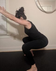
- Excessive lordosis—An anterior pelvic tilt (APT) is most commonly associated with LBHCD, but, accompanied with some of the above signs, can also signal LED. Similar to (and often occuring along with) excessive forward lean, the hip flexors are short: rectus femoris, anterior adductors and TFL. Glute max and medius are weak.
OHSA with Modification
Here’s how to determine if any of the above problems are more related to LED or LBHCD.
Have the client perform the overhead squat again, but this time with the heels raised about two inches on a half foam roll (flat side down), block or riser. Doing this will take any short plantarflexors out of the equation. If any of the noted signs vanish with this modification, then it can be attributed to LED. If it remains, the problem is more likely to be associated with LBHCD.
What To Do With Noted Signs of Dysfunction
Familiarizing yourself with anatomy and joint actions is crucial for moving forward with corrective exercise. The most sensible and conservative thing to start with is to perform myofasical release of the overactive muscles and then lengthen them with stretching.
Simultaneously, the weak musculature should be activated and strengthened. These are steps to take before integrative exercises can be implemented and OHSA re-assessed to note any progress.
Stay tuned for additional blogs analyzing signs of LPHCD and Upper Body Dysfunction (UBD).
*The above concepts are directly adapted from the Brookbush Institute and its Human Movement Specialist certification which I am currently pursuing.